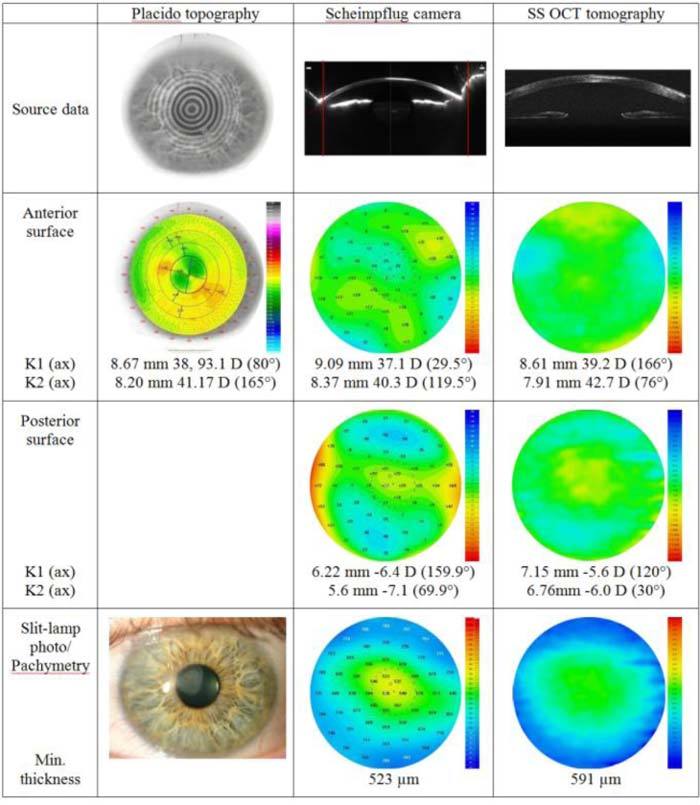
Fig. 5.

Qualitative evaluation of a cornea with superficial postinfectious scar with three different instruments. K1, K2, central keratometry readings. The red lines on Scheimpflug images correspond to the lateral size of cross-sectional images for SS OCT.
