Fig. 6.

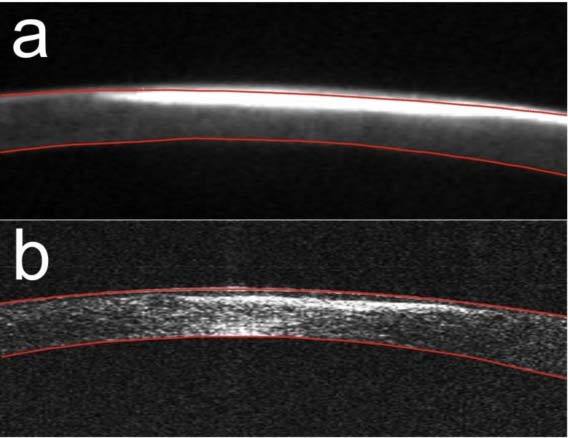
Cross-sectional images of the cornea with superficial postinfectious scar: (a) Scheimpflug image measured by Pentacam HR; (b) SS OCT cross-sectional image. Red lines delineate the segmented corneal boundaries.

Cross-sectional images of the cornea with superficial postinfectious scar: (a) Scheimpflug image measured by Pentacam HR; (b) SS OCT cross-sectional image. Red lines delineate the segmented corneal boundaries.