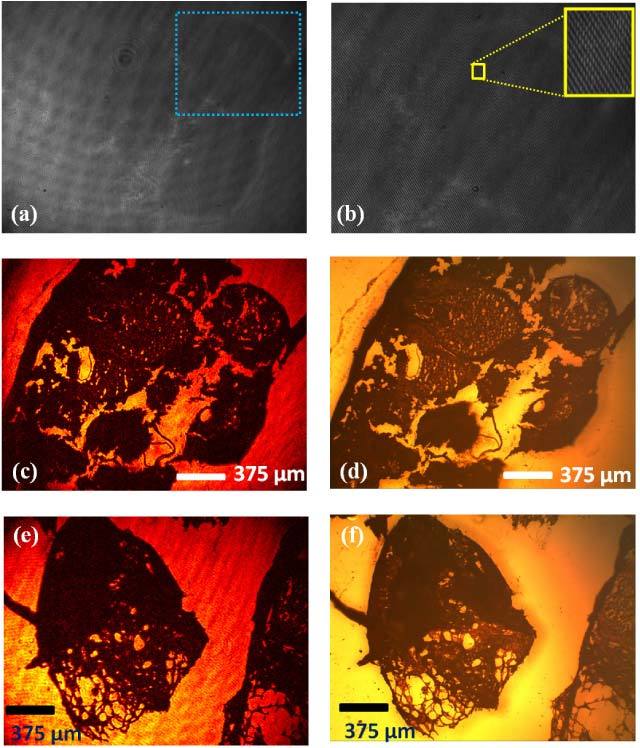
Fig. 4.

Reflection imaging of a histopathology slide corresponding to skin tissue using lensfree off-axis holography. (a) The raw off-axis reflection hologram of skin tissue. (b) The digitally zoomed hologram region specified with the blue rectangle in Fig. 4(a) is shown. The corresponding reconstructed amplitude reflection image is shown in (c). Conventional reflection mode microscope image of the same specimen using a 4X objective lens (NA: 0.1) is also shown in (d) for comparison purposes. Note that due to their limited FOV, higher magnification objective lenses would not be able to capture the same comparison image. (e) same as in (c) except for a different region of interest. (f) provides a conventional reflection mode microscope image of the same specimen for comparison.
