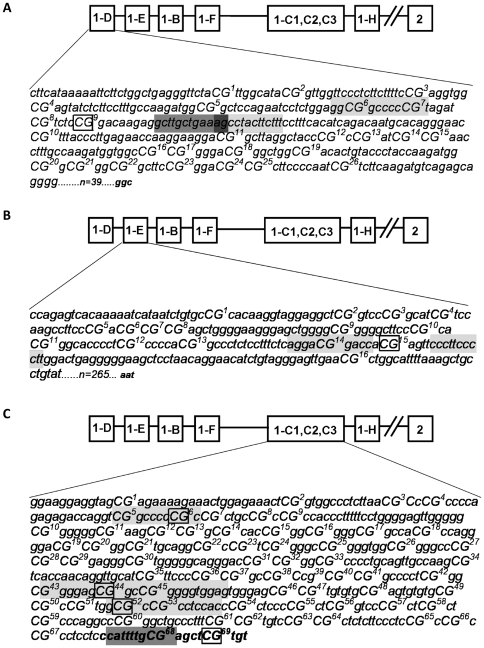
Figure 3. CpG methylation in the GR 1C, 1D, and 1E promoter regions in DMS 79 cells.
Bisulphite sequencing was performed on extracted genomic DNA from DMS 79 cells incubated with 5′Azadeoxycytidine for 5 days (untreated as control). The original sequence (obtained from GENOME Browser) was compared to bisulfite treated DNA from the control and 5′Azadeoxycytidine treated samples. Bold, open boxes highlight specific methylated CpGs, which showed reversal of methylation after 5′Azadeoxycytidine incubation. The potential transcription factor binding sites in close proximity to the methylated sites are indicated by grey shading for Sp1 binding site and dark grey shading for C/EBPβ binding site. (A) CpG methylation in the GR 1D promoter region in DMS 79 cells. Individual CpGs are numbered and in capitals. (B) CpG methylation in a region of the GR promoter 1E from DMS 79 genomic DNA. Individual CpGs are numbered and in capitals. (C) Genomic DNA from a region of the GR promoter 1C from DMS 79 cells. Individual CpGs are numbered and in capitals. Italic text indicates the start of exon 1C.

