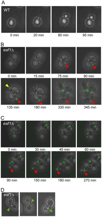
Figure 3. eaf1Δ cells have defects in septin dynamics.
Time lapse imaging of cells expressing Cdc11-GFP in the presence (WT, YKB1312) or absence of EAF1 (eaf1Δ, YKB1310). Cells were grown to mid-log phase in YPD supplemented with adenine at 25°C, and transferred onto synthetic complete agarose pads for imaging at room temperature. Images were collected at five or fifteen minute intervals for wild type and eaf1Δ cells, respectively. Representative images of cells over time are shown in panel (A) for wild type cells and panels (B–C) for eaf1Δ cells. Red arrows point to septins mislocalized into the bud. The yellow arrow points out initially mislocalized septins that re-localize to the bud neck, followed cell division. (D) Images of three different eaf1Δ cells exhibiting septins misorganized to one side of the neck. Quantification of live cell imaging is presented in Table 1.

