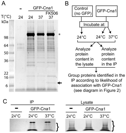
Figure 1. Mass spectrometry (MS) analysis of calcineurin-associated proteins.
A. A Coomassie blue-stained gel shows that the GFP-Trap resin exhibits a very low affinity towards proteins present in the control lysate, whereas GFP-Cna1 (star) associates with a large number of proteins. An arrow indicates a protein that likely corresponds to FKBP12 in a sample treated with FK506. B. Scheme depicting general proteomics approach/strategy. C. Coomassie blue-stained gels of the samples that were precipitated using the GFP-Trap resin (left) and the cell lysates analyzed for the protein content as a control (right). For each gel, an area that was excised from the gel for the subsequent MS analysis is depicted (brackets).

