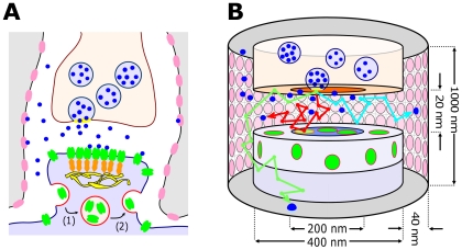
Figure 1. Representation of the synapse dynamics.
(A) Sketch of an excitatory synapse consisting of the presynaptic terminal where vesicles are released, and the postsynaptic element where glutamate receptors are located. The synapse is surrounded by astroglial processes containing glutamate transporters (GLTs). Presynaptic vesicle fusion occurs at randomly selected locations, released glutamate (blue) diffuses in the cleft and binds to AMPARs (green) or GLTs (pink). AMPARs diffuse between the PSD, where they can attach to scaffolding molecules (orange) and the extrasynaptic regions, where they can undergo endocytosis (1) and exocytosis (2), maintaining the number of AMPARs at the post-synaptic terminal. (B) Two co-axial cylinders represent the pre- and postsynaptic terminal, forming a gap which represents the synaptic cleft. AMPARs (green) are distributed inside and outside the PSD. The trajectory of a glutamate molecule as illustrated by red, blue or green arrows corresponds to binding to AMPARs, GLTs or diffusing away from the cleft (at 500 nm), respectively.

