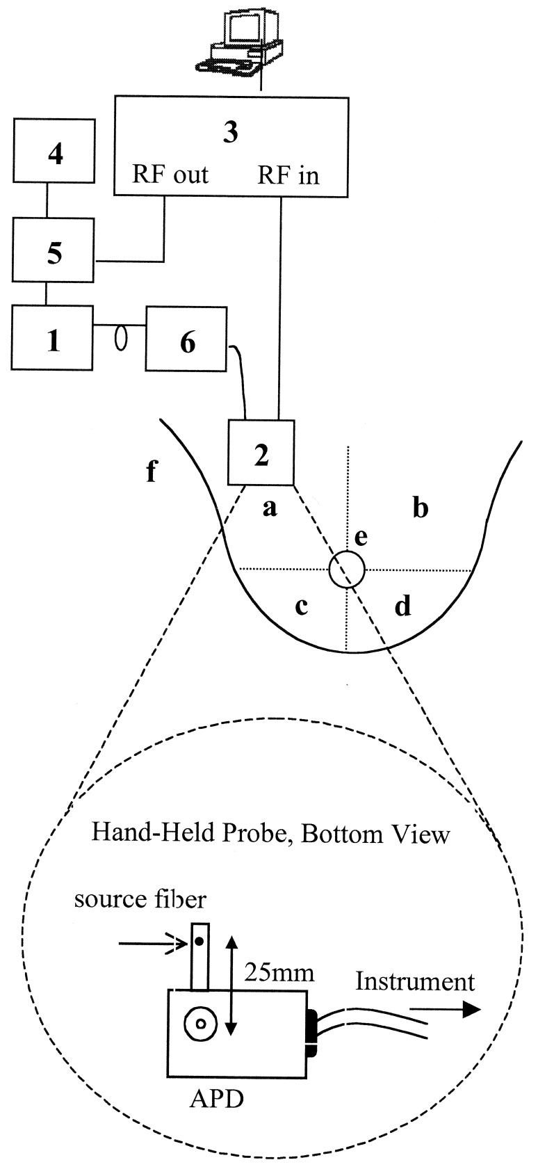
Figure 1.
Schematic drawing of FDPM instrument, hand-held probe, and measurement map of healthy subjects. The components of the instrument are: diode lasers (box 1), avalanche photodiode (box 2), network analyzer (box 3), DC current source (box 4), bias T (box 5), and optical switch (box 6). See text for detailed description. The breast is divided into four quadrants: upper outer (a), upper inner (b), lower outer (c), and lower inner (d). FDPM measurements are made in each quadrant, on the areolar border (e), and on the glandular tail (f) that extends into the axilla.

