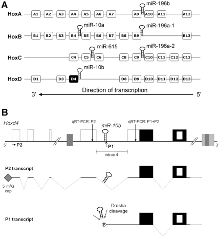
Figure 1. Hox genes and their associated microRNAs.
(A) A schematic of the four mammalian Hox clusters (HoxA through HoxD) and the position of the microRNAs embedded within them [20]. The indicated direction of transcription applies to all four Hox clusters. (B) A schematic showing the genomic organization of the mouse Hoxd4 gene. Empty boxes indicate non-coding exons and black boxes indicate coding exons. The nested white box indicates the homeobox within the second coding exon. Grey boxes show regulatory elements. P2 denotes the upstream promoter and P1 denotes the putative downstream promoter. miR-10b is found directly upstream of the P1 promoter, in intron 4 of the P2 transcript. Dotted lines indicate spliced introns. The grey diamond at the 5′ end of the P2 transcript denotes the 5′ 7-methylguanosine (m7G) cap and the circled “P” at the 5′ end of the P1 transcript indicates a 5′ phosphate. The positions of two 200 bp regions amplified by qRT-PCR (qRT-PCR∶P2 and qRT-PCR∶P1+P2) are indicated by vertical arrows (see figure 2).

