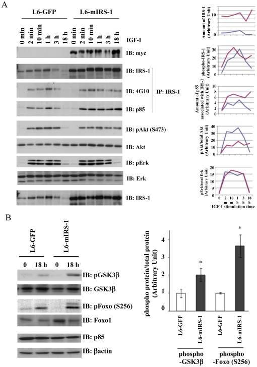
Figure 3. Effects of IRS-1 constitutive expression on IGF-I signal activation in L6 myoblasts.
A, B: L6-GFP cells or L6-mIRS1 cells were serum starved for 8 h, followed by stimulation with IGF-I (100 ng/ml) for indicated time (0 min, 2 min, 10 min, 1 hour, 3 hour and 18 hour). Cells were harvested and lysed in RIPA buffer. One hundred µg of cell lysates were immunoprecipitated with anti-IRS-1 antibody (IP). Ten µg of total cell lysates or immunoprecipitates were separated with SDS-PAGE and immunoblotted with indicated antibodies (IB). Bands were quantified from each blot by NIH Image J software. Protein amount of IRS-1, p85 associated with IRS-1, phosphorylated IRS-1 or phosphorylated proteins over total proteins (pAkt/Akt, pErk/Erk, pGSK3β/GSK3β and pFoxo1/Foxo1) was calculated and the values were shown in the graphs. B: Values are the mean ± SEM of three different experiments and expressed as relative to data in insulin-stimulated L6-GFP cells. *, the difference between L6-GFP cells and L6-mIRS1 is significant with p<0.05.

