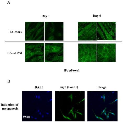
Figure 6. Foxo1 localization in L6 and satellite cells.
A: L6-mock or L6-mIRS1 was incubated in DMEM containing 2% FBS for 18 h and 6 days. Cells were immunostained with anti-Foxo1 antibody. Foxo1 localization is shown. B: Satellite cells were separated from the rat soleus muscle and incubated in DMEM containing 20% FBS. Plasmid expressing myc-tagged Foxo1 H215R mutant was transfected into satellite cells. One day after transfection, muscle differentiation was induced by changing medium to 2% FBS. One day after induction of differentiation, cells were fixed, permeabilized and immunostained with anti-myc antibody. DAPI staining was shown in blue. Myc staining (Foxo1) is shown in green.

