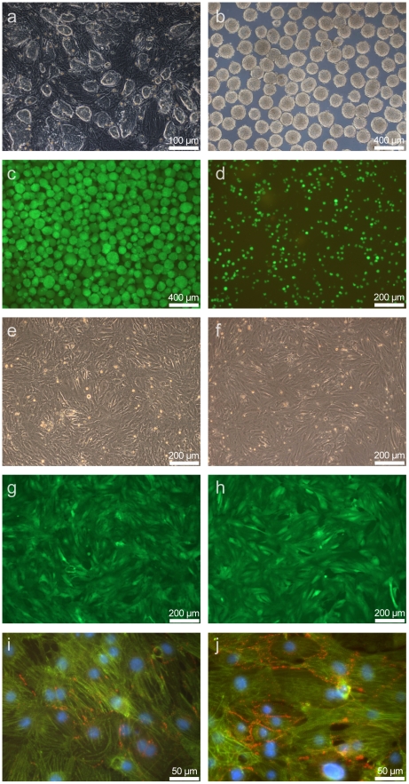
Figure 1. Differentiation and maturation of undifferentiated ES cells to Cor.At® cardiomyocytes.
a) Undifferentiated mouse ES cells (clone αPIG44) on feeder cells. b) Mouse ES cells aggregated into EBs at day 3 after initiation of differentiation. c) Mouse ES cell derived Cor.At® cardiomyocytes after 12 days of differentiation and 3 days of puromycin treatment, before dissociation. d) Mouse ES cell derived Cor.At® cardiomyocytes after 12 days of differentiation and 3 days of puromycin treatment, after dissociation (probe Cor.At® cardiomyocytes 12 days). e,g,i) Cor.At® cardiomyocytes after additional 7 days of culture (probe Cor.At® cardiomyocytes 19 days): e) transmission, g) same region EGFP fluorescense indicating differentiation to cardiomyocytes, i) overlay of immunostainings: blue = nucleus staining with DAPI, green = α-actinin (structured) and GFP, red = connexin 43. f, h, j) Cor.At® cardiomyocytes after additional 7 days of culture (probe CorAt 26 days): e) transmission, g) same region GFP fluorescense indicating differentiation to cardiomyocytes, i) overlay of immunostainings: blue = nucleus staining with DAPI, green = α-actinin (structured) and GFP, red = connexin 43.

