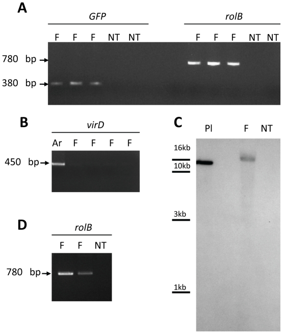
Figure 2. Detection of transgenes by PCR and RT-PCR.
A-B PCR analysis of genomic DNA isolated from P. japonicum fluorescent hairy roots (F) and non-transformed tissues (NT). A. Amplification of GFP and rolB fragments with expected sizes (380 bp and 780 bp, respectively). B. No amplification of virD1 fragment (450 bp) in fluorescent tissues (F), as positive control a diluted ATCC15834 bacterial suspension (Ar) was used. C. Southern blot of genomic DNA extracted from fluorescent (F) and non-transformed (NT) P. japonicum roots. DNA was digested with EcoRI. The positive control (Pl) corresponds to linearised pBCR101 plasmid (30 ng). D. RT-PCR analysis of the rolB gene using total RNAs extracted from P. japonicum fluorescent roots (F) and non-transformed tissues (NT).

