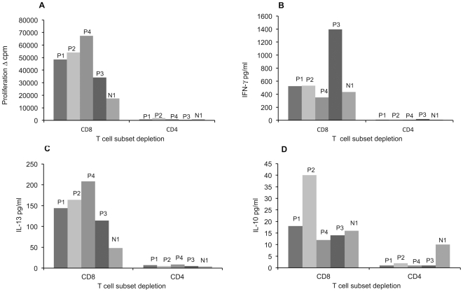
Figure 1. MCPyV VP1–specific T cell responses of MCPyV seropositive individuals after T cell subset depletion.
PBMC of four MCPyV seropositive (P1 to P4) and one seronegative subject with strong MCPyV-specific CMI (N1) were depleted of either CD4+ or CD8+ T cells and stimulated with MCPyV VP1-VLPs (2.5 µg/ml). Proliferation (panel A) and cytokine (IFN-γ, IL-13 and IL-10) responses (panel B, C, D) were studied by thymidine incorporation and ELISA, respectively.

