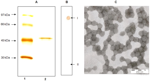
Figure 3. Characterization of MCPyV VP1 antigens.
Silver staining of capsid protein (panel A) in 10% SDS PAGE. Lane 1: molecular weight markers, lane 2: MCPyV VP1 capsid antigen. Dot blotting (panel B) for MCPyV antigen, studied with MCPyV-IgG positive (I) and negative (II) sera. Electron microscopy of sterile MCPyV particles (panel C) purified by caesium chloride density gradient ultracentrifugation, with 200 nm scale bar shown.

