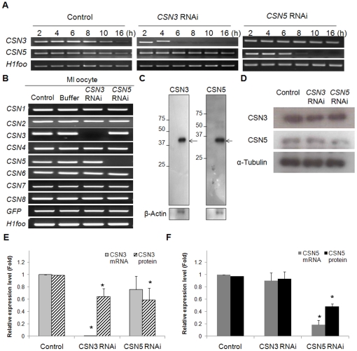
Figure 2. Efficiency of RNAi-mediated knockdown of CSN3 and CSN5.
(A) Determination of the critical time point for complete knockdown of CSN3 and CSN5 after RNAi. Oocytes were collected every 2 h after RNAi, and CSN3 and CSN5 mRNA was assessed by RT-PCR. (B) Specific suppression of CSN3 or CSN5 mRNA by RNAi. The other CSN subunits were not affected by RNAi treatment. H1foo was used as an internal control of oocytes, and exogenous GFP mRNA was used as an external control. (C) Western blot analysis of CSN3 and CSN5. The blot was incubated with each antibody using 100 oocytes. Numbers on the left side of the band indicate the sizes (kDa) of the protein markers, while arrows indicate the specific proteins. β-Actin was used as a loading control. (D) Protein knockdown after RNAi. Levels of CSN3 and CSN5 protein in RNAi-treated oocytes were determined using Western blot analysis. Proteins were extracted from 100 MI oocytes for each lane. (E, F) Bar graphs show the relative mRNA and protein levels after RNAi. The mRNA level was calculated with quantitative real-time RT-PCR using single-equivalent oocyte cDNA, while protein level was calculated by measuring the density and area of the bands. The mRNA and protein levels are presented relative to those of control oocytes. Asterisks indicate statistically significant differences compared to that of control or buffer group (p<0.05).

