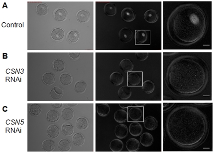
Figure 4. Down-regulation of CSN3 or CSN5 caused meiotic spindle disassembly.
Spindle structure was observed noninvasively using Polscope microscopy. (A) Control MI oocytes cultured for 8 h showed normal barrel-shaped spindles. (B, C) After RNAi treatment followed by 16 h culture, oocytes were arrested at the MI stage and showed no spindle structure. Left panel, bright field; middle panels, dark field; right panels, magnified view of boxed area from the middle panels. Original magnifications ×200 (Left and middle panels). Bar s = 20 µm.

