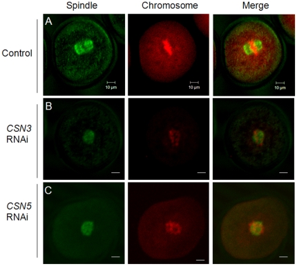
Figure 6. Immunofluorescence staining of the spindle and chromosomes.
Spindles were stained with α-Tubulin antibody (green) and chromosomes were counterstained with propidium iodide (red). (A) Control MI oocytes cultured for 8 h. (B, C) RNAi-mediated knockdown of CSN3 (B) and CSN5 (C) arrested oocytes at the MI stage and the oocytes showed abnormally aggregated spindle and chromosomes. Left panel, spindle structure; middle panel, chromosome; right panel, merged image. Bar = 10 µm.

