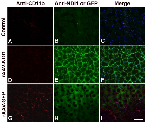
Figure 3. Representative images of macrophage staining of tissue sections from rat muscles.
Rats were injected with PBS, rAAV-GFP, or rAAV-NDI1 in the skeletal muscles and muscle samples were collected 1 month post-injection. Coronal sections of the muscle from the animals injected with PBS (Control), rAAV-NDI1 or rAAV-GFP were stained with anti-CD11b antibody (A, D, and G, red) or with anti-Ndi1 antibody (E, green). Muscle sections from the rats injected with rAAV-GFP were examined for the fluorescence of the GFP protein (H, green). A green channel image for the control (B) is also shown. Merged images of the red and green channels are also shown (C, F, and I) Scale bar = 75 µm.

