Fig. 3.
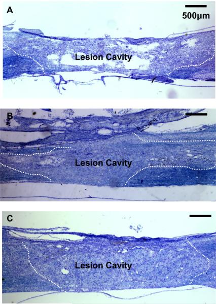
Nissl-Myelin staining at the lesion site. Dotted lines indicate lesion border.
A) Longitudinal section of spinal cord from OP-Control rat. The central lesion consisted cyst formation and cavitation.
B) Longitudinal section of spinal cord in Nissl-Myelin staining shows survival of NRP/GRP transplanted into the injured spinal cord at 8 weeks post-transplantation. Dotted line indicates graft-host interface. The cavity was largely filled with transplanted cells, and few cysts were apparent.
C) Longitudinal section of spinal cord in Nissl-Myelin staining shows that transplanted NRP/GRP survived and combined treatment of NRP/GRP and NBQX protected more host tissue than single treatment of NRP/GRP.

