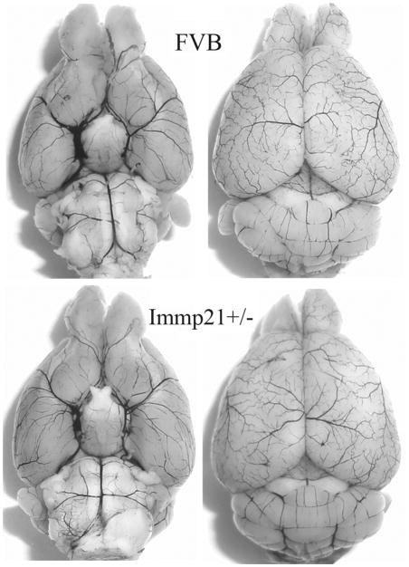
Fig. 2.
Cerebral vasculature. Mice (n=3 each group) were perfused with Indian black ink to determine whether there were vascular abnormalities in the Immp2+/− mice. The Circle of Willis, anterior cerebral arteries, middle cerebral arteries, and posterior arteries all appear normal as compared with those in WT (FVB) controls.

