
FIG. 1.
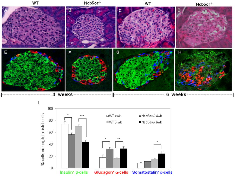
(A–D) H & E staining and (E–H) confocal immunofluorescence analyses against insulin (β-cells, green), glucagon (α-cells, red) and somatostatin (δ-cells, blue) of representative islet sections from Ncb5or−/− and WT mice at age 4 and 6 weeks. (I) Average percentages of each cell type (n=3 mice per group; 5 islets per mouse section). *, p<0.05. **, p<0.01. ***, p<0.005.