
FIG. 4.
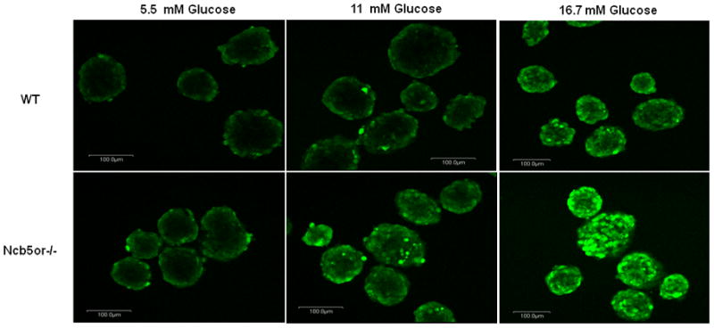
DCF staining of higher H2O2 levels in Ncb5or−/− islet β-cells. Islets from Ncb5or−/− and WT mice at age 5 weeks were incubated with 5.5, 11 and 16.7 mM glucose for one hour prior to DCF staining. A total of three pairs (n=3) were analyzed and representative images were shown. Signals in peripheral are likely resulted from physical barrier for penetration of dye.