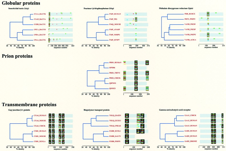
Figure 1.
The localization of TM helices in globular, transmembrane, and prion proteins. For comparison, 6–6 homologs of three TM and three globular proteins that have a similar relative similarity dendogram to that of the selected six prion proteins were selected (proteins are given by SWISS-PROT accession no. or ID). For each protein, TM helices are predicted by four prediction methods and are shown by color coding as follows: TOPPRED (green), DAS (red), PHDhtm (blue), and HMMTOP (yellow). A TM region (defined as in the legend of Table 2) is boxed if predicted by three (gray) or four (black) methods. Please note that there are only 11 globular proteins of 523 for which at least three methods predict a TM region (cf. Table 2); only three could be found with a relative similarity dendogram as shown.

