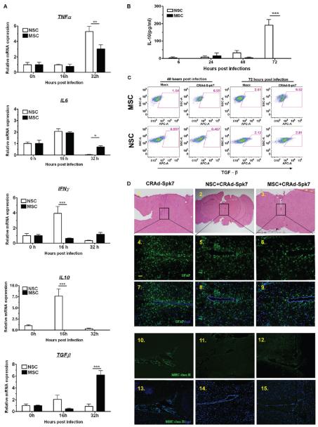
Figure 5.
Immunological consequences of oncolytic adenovirus loading into stem cells. (A) Analysis of cytokine expression postinfection with 10 IU of CRAd-S-pk7 virus per stem cells by qRT-PCR. Levels of expression were expressed relative to uninfected control for each stem cell line at 0 h; *P < 0.05, **P < 0.005, ***P < 0.0005 significant. (B) Analysis of IL-10 cytokine expression in stem cells infected with CRAd-S-pk7 (10 IU/cell). Supernatant from the infected stem cell culture was harvested at 6, 24, 48 and 72 h postinfection, and IL-10 expression was measured by ELISA; ***p <0.0005. (C) Analysis of TGF-β positive stem cells by FACS at 48 and 72 h postinfection with CRAd-S-pk7 (10 IU/cell). (D) Histological features of brains from mice injected with CRAd-S-pk7 virus alone (2.5 × 10⁷ iu/injection) or virus loaded into stem cells (5 × 10⁵ cells loaded with 50 IU of CRAd-S-pk7 per NSCs) harvested at day 14 postinjection. (1–3) Image of H&E staining assessing neuronal toxicity postinjection. Representative image of sections of the same animal brain as H&E sections stained with antiglial fibrillary acidic protein (GFAP) (4–9) and anti-mouse MHC class II antibody (10–15); bar, 50 μm.

