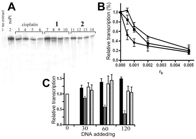
Figure 6.
Inhibition of RNA polymerase II transcription by DNA adducts of complexes 1, 2 and cisplatin. (A) Autoradiogram of the 8% PAA/8 M urea denaturing gel. Lanes: 1, unmodified substrate, no extract added; 2, control, unplatinated pCMV-Gluc substrate; 3, 4, 5 and 6, pCMV-Gluc substrate modified with cisplatin at rb = 5×10⁻⁴, 1×10⁻³, 2×10⁻³ and 5×10⁻³, respectively; 7, 8, 9 and 10, pCMV-Gluc substrate modified by complex 1 at rb = 5×10⁻⁴, 1×10⁻³, 2×10⁻³ and 5×10⁻³, respectively; 11, 12, 13 and 14, pCMV-Gluc substrate modified by complex 2 at rb = 5×10⁻⁴, 1×10⁻³, 2×10⁻³ and 5×10⁻³, respectively. B. Quantitative assessment. The relative transcription was assed as follows: the amount of full length transcript at each rb was quantified (in % of total radioactivity in the lane) and calculated as the percentage of that generated by the control, undamaged template. Data represent results of two independent experiments and are expressed as mean percentages ±SEM. (■) cisplatin; (▲) 1; (●) 2. C. Inhibition of RNA polymerase II transcription by the addition of increasing amount of exogenously platinated pUC19 DNA. The amount of full-length transcript in each lane is expressed as a mean fraction (±SEM) of that generated in the absence of exogenously added DNA (white bar). Black bars: transcription of undamaged DNA; gray bars: transcription of cisplatin modified DNA; light gray bars: transcription of DNA modified with 1; hatched bars: transcription of DNA modified with 2.

