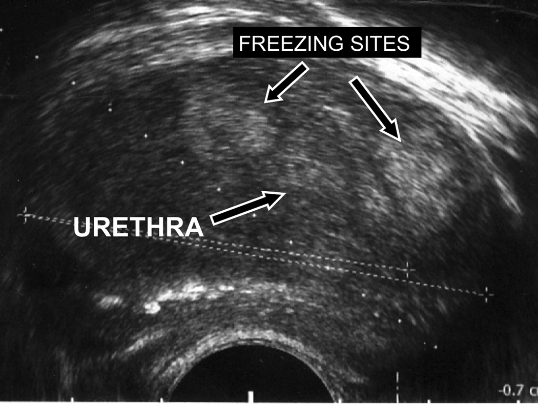
Figure 4. Post treatment TRUS.
Axial transrectal ultrasound of the prostate gland using a 7.5 MHz biplanar endorectal ultrasound probe. Two echogenic foci are present in the gland antero-lateral to the urethra, and appear similar in configuration to the sites of cryoablation seen on the MR images during freezing, as well as the areas of devitilization seen on the subtracted post-contrast images.

