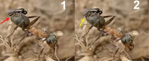
Figure 6.
Insertion of the ovipositor by Elasmosoma luxemburgense. 1 the red arrow shows the wasp’s metasoma separated from the ant’s metasoma 2 the yellow arrow shows the metasoma of the parasitoid and of the ant joined during insertion of the wasp’s ovipositor. The fore legs have now advanced their position towards the posterior margin of the first gastral segment.

