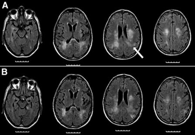
Figure 1.

MRI, FLAIR: (A) At admission. Confluent hyperintense areas in T2/FLAIR in the periventricular and subcortical white matter, extending to the right parietal cortex and basal ganglia. The lesions were mildly hypointense in T1 and showed discrete gadolinium enhancement. Stereotactic biopsy site (arrow). (B) Two weeks after treatment.
