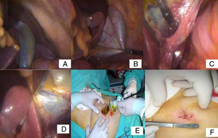
Figure 3.

(A) The liver and the gallbladder are seen in the left upper quadrant of the abdomen. (B) The gallbladder was suspended from the fundus to the anterior wall of the abdomen. (C) The cystic duct and artery were identified dissecting Calot’s triangle above Rouviere’s sulcus. The cystic duct and artery were clipped and divided. (D) The gallbladder was excised retrogradely from the liver bed. (E) The gallbladder was extracted from the umbilical incision. (F) The skin was closed subcutaneously.
