Figure 7.
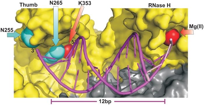
A crystal structure of RT in complex with a dsDNA substrate (PDB 3KK2); only a portion of the dsDNA substrate that extends 12 bp from the RNase H active site is shown to approximate the position of the stem of RT1t49(−5), the positions equivalent to the 5′- and 3′-ends of the stem are indicated. The figure illustrates how the stem of RT1t49(−5) can protect K353. Residues N255 and N265 that play a role in recognition of RT1t49 are shown (cyan). N255 is not contacted by the nucleotides in the 12 bp shown, and N265 is in proximity to the 5′-end of the portion of the template stand that is shown.

