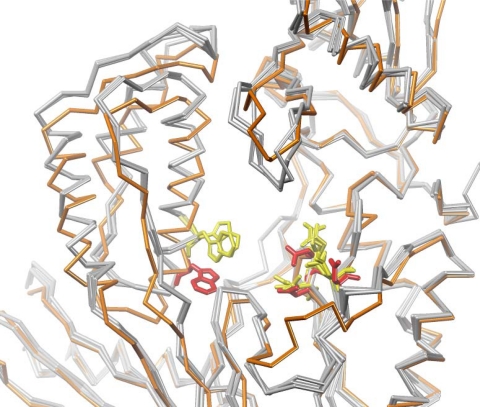
Figure 9.
Detailed view of the DAVP-1 binding pocket showing a superposition of the cα backbone in unliganded HIV-1 RT structures (in grey, PDB 1DLO, 1QE1 and 1HQE) and in RT bound to DAVP-1 (in orange, PDB 3ITH and 3ISN). Residues W266, D110, D185 and D186 are shown in yellow in unliganded RT structures and in red in the RT/DAVP-1 complex.

