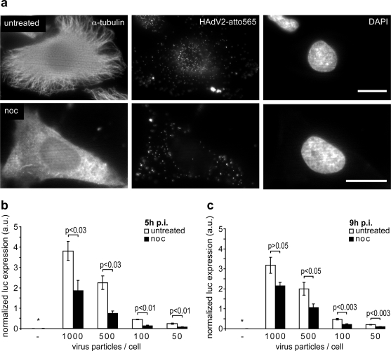
Figure 1.
Adenovirus infection requires intact microtubules. (a) HeLa cells were kept in the presence or absence of nocodazole for 30 min, inoculated with 0.5 μg HAdV2-atto565 and incubated at 4 °C for 30 min. Temperature was shifted to 37 °C and infection proceeded for 90 min, cells were fixed with PHEMO fixative [42], processed for DAPI and immunostaining against alpha-tubulin and imaged by wide field fluorescence microscopy. Scale bars 20 μm. (b, c) HeLa cells were continuously treated or left untreated with nocodazole in Hoechst containing medium, infected with the indicated physical virus particle to cell ratios for 5 h (b) or 9 h (c), lysed and luciferase expression was quantified. Before cell lyses and in the last 15 min of infection, cells were imaged with an ImageXpress Micro Imaging Station (Molecular Devices) in the Hoechst channel. Luciferase expression was normalized to the number of nuclei detected in the 16 sites imaged per well. An asterisk indicates samples with luciferase expression below the detection limit. The means ± SEM of 4 (b) or 3 (c) independent experiments are shown. The two-tailed Student's t test was used to determine p-values.

