Figure 1.
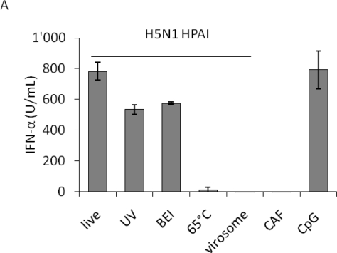
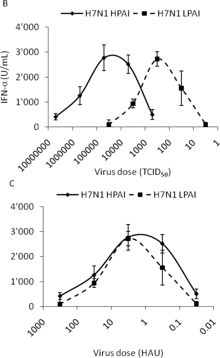
IFN-α responses of plasmacytoid dendritic cells (pDC): impact of virus inactivation, as well as virus-dose dependency using either HAU or TCID50 to quantify influenza A virus (IAV). Porcine pDC were enriched from fresh blood, plated and stimulated. IFN-α in the supernatant was quantified by ELISA after 24 h. (A) pDC were stimulated with live, UV-, 2-bromoethylamine hydrobromide (BEI)- or 65 °C heat-inactivated H5N1 virus (H5N1 TT05, see Table 1) or with H5N1-derived virosomes at a dose of 40 HAU. Allantoic fluid (CAF) or CpG (10 μg) were used as controls. Error bars represent standard deviations of triplicate ELISA samples. (B and C) pDC were stimulated with a pair of HP- and LPAIV H7N1 (H7N1 TI99 and OI00, see Table 1) at various doses. Error bars represent standard deviations of triplicate samples.


