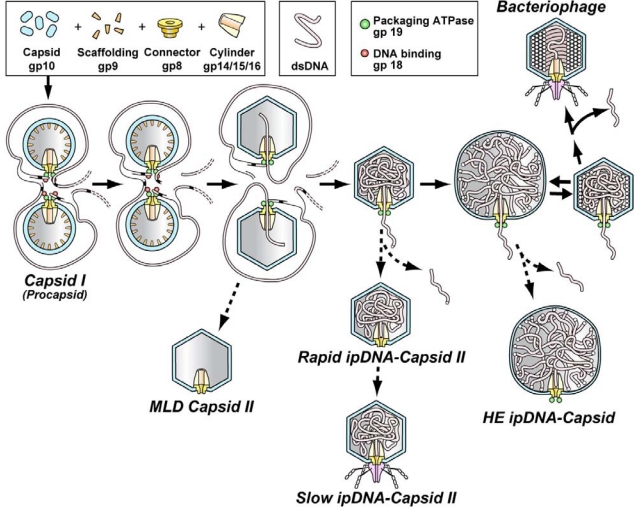
Figure 1.
The DNA packaging pathway of the related bacteriophages T3 and T7 (adapted from [30]). The solid arrows indicate the proposed productive pathway in an infected cell. The dashed arrows indicate the pathways for generating the motor-related particles that have been observed by fractionation and characterization. Duplication of the early stages represents cooperativity detected by single-molecule fluorescence microscopy [93]. The legend at the top indicates the color-coding of both the DNA molecule and the various proteins.

