Figure 3.
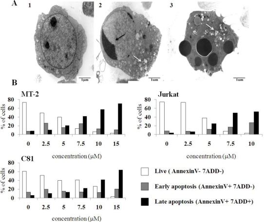
C7a induces morphological and phenotypic alterations compatible with apoptosis in human leukemia cell lines. (A) Transmission electron microscopy. (1) HL-60 cells cultivated in complete RPMI medium for 6 h; (2 and 3) HL-60 cells cultivated in presence of 2 μM C7a for 7 h. Black arrows: nuclear condensation; White arrows: vacuoles. This assay was repeated twice, and representative images are shown; (B) Membrane phosphatidylserine expression. HTLV-1 infected MT-2 and C81, and uninfected Jurkat cell lines (1 × 10⁶) were incubated in the presence of C7a at the indicated concentrations for 3 h at 37 °C. After staining with PE-Annexin V and 7-AAD, cells were analyzed by flow cytometry, as described in Material and Methods. A representative of two independent experiments is shown.

