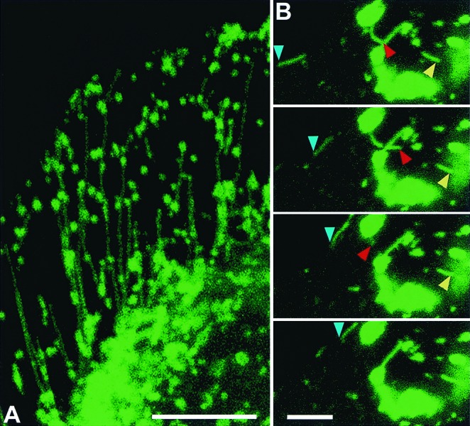
Figure 2.

NPC1-GFP traffic via late endosomal tubules in CHO cells. (A) CT60 CHO cells expressing NPC1-GFP (green) were fixed and visualized by confocal microscopy (the cell shown is the one in Fig. 1 E). (B) Live-cell, confocal-scanning time sequence of NPC1-GFP-containing tubules, showing rapid motion of three NPC1-GFP tubules with variable rates. Blue arrowheads mark tubular trafficking along the periphery of the cell. Red arrowheads mark tubular extension and retraction. Yellow arrowheads mark tubular trafficking among vesicles. The time interval between images is 2 s. All images are confocal. [Bar = 10 μm (A) or 5 μm (B).] View dynamics of late endosomal tubular trafficking of NPC-1 GFP in Movies 1 and 2.
