Figure 3.

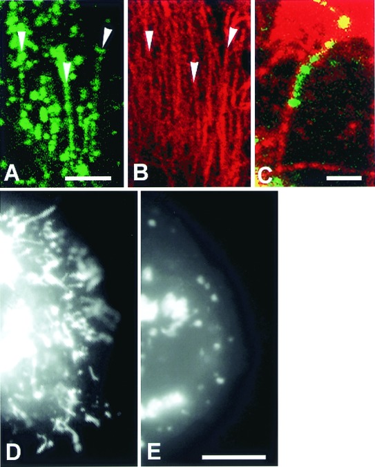
NPC1-GFP-containing late endosomal tubular trafficking is microtubule dependent. (A–E) CT60 CHO cells transfected with NPC1-GFP. Confocal images of fixed cells show NPC1-GFP tubules (A, green) aligned along microtubules (B, labeled with anti-α-tubulin, red). Arrowheads in A and B indicate areas of obvious alignment. (C) A merged image shows a NPC1-GFP tubule (green) aligned along a microtubule (red). (D and E) Digital images of living CT60 CHO cells. (D) A CT60 CHO cell cleared of cholesterol contains many NPC1-GFP-containing late endosomal tubules. (E) A CT60 cell incubated with nocodazole for 70 min. NPC1-GFP-containing late endosomal tubules are not present in cells in which microtubules are depolymerized by nocodazole treatment. A–C are confocal images, D and E are digital epifluorescence images. [Bar = 10 μm (A and B), 2 μm (C), and 10 μm (D and E).]
