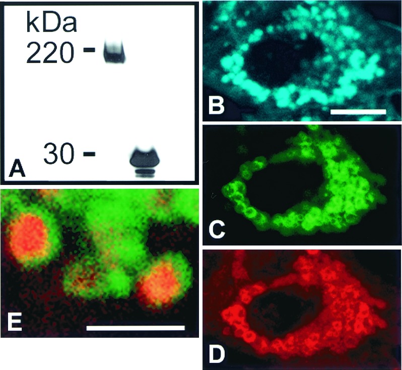
Figure 6.

Late endocytic trafficking is blocked by cholesterol accumulation in CT60 CHO cells expressing SSD-NPC1-GFP. (A) Western blot analysis with anti-GFP antibody of total solubilized protein from SSD-NPC1-GFP-expressing WT CHO cells (Left) and EGFP-expressing WT CHO cells (Right). The predicted molecular mass of GFP (27 kDa) combined with glycosylated SSD-NPC1 (170 kDa) is comparable to the 200-kDa band (Left). (B–E) CT 60 CHO cells expressing SSD-NPC1-GFP fail to clear cholesterol. Seventy-two hours after transfection, cells were fixed, stained, and labeled with filipin (B, blue) to visualize cholesterol by confocal microscopy. SSD-NPC1-GFP (C, green) was also detected by indirect immunofluorescence (D, red). (E) Cholesterol-laden lysosomes in an SSD-NPC1-GFP transfected CT60 CHO cell are labeled by endocytic uptake of rhodamine-dextran. In this merged image, SSD-NPC1-GFP (green) is present as rings at the periphery of rhodamine-dextran (red) in the cholesterol-laden core of lysosomes. B–E are confocal images. [Bar = 10 μm (B–D) and 2 μm (E).]
