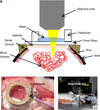
Figure 1.
The experimental setup. (A) Sagittal view of the entire setup. (B) After a craniotomy, the single-element piezoelectric transducer, attached to a glass cover slip, was secured with cyanoacrylate glue and dental cement. The ring transducer also served as water well for water-immersion objective lens. (C) Animal is secured on streotaxic stage for two-photon imaging.

