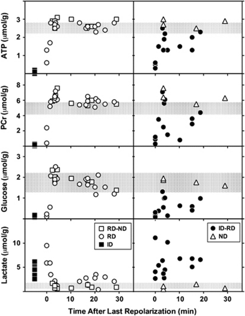
Figure 3.
Metabolite changes in response to depolarization. Tissue samples from sites identified in Figure 2 were obtained from brains frozen in situ after 1 hour middle cerebral artery (MCA) occlusion, during or at intervals up to 30 minutes after a recurrent depolarization (RD). Locations undergoing persistent ischemic depolarization (ID, filled squares, n=9) exhibited loss of ATP, phosphocreatine (PCr) and glucose, and markedly elevated lactate, relative to corresponding tissue from sham-operated animals (shaded bars, mean±s.d.). RD (open circles, n=17) resulted in profound but transient metabolic responses, tending to overshoot control values 3 to 5 minutes after repolarization. Increases in PCr were also observed during this interval in ipsilateral cortex that had not exhibited depolarization (ND, open triangles, n=4), and at adjacent locations (RD–ND, open squares, n=9). Thereafter ATP and PCr levels remained stable, whereas glucose declined slightly and lactate increased modestly above control levels. Persistent, intermediate changes in metabolite levels were only observed in regions adjacent to those undergoing sustained depolarization (ID–RD, closed circles, n=13).

