Figure 2.
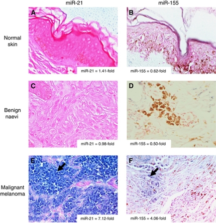
In situ hybridisation for miR-21 and miR-155 in (A and B) normal skin (200 × ), (C and D) benign naevi (400 × ) and (E and F) malignant melanoma (400 × ). Specific staining for miR of interest is blue (arrow) and counterstain is pink, some samples contain melanin pigment (brown coloration). Validation of miR-21 and miR-155 expression as determined by real-time PCR is indicated in the corner of each panel. Data shown are representative of n=4 samples for each tissue type.

