Figure 2.
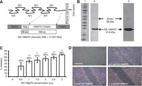
The truncated form of CD9-P1, GS-168AT2, inhibits dose-dependently in vitro hEC proliferation and migration. (A) Schematic representation of the structure of CD9-P1, the cloned truncated CD9-P1 domain, GS-168AT2, and the produced Tag-recombinant protein. (B) SDS–PAGE imprints (lane 1, molecular mass marker; lane 2 purified GS-168AT2) and the corresponding WB (lane 3, using 229 mAb) of the purified GS-168AT2 used for the all subsequent experiments. (C) Dose-response curve of the effects of GS168AT2 on the proliferation of hEC. Results were expressed as percentage of control±s.e. (n=4). Statistical significance between the control and the different doses of GS-168AT2 were calculated with two-tailed Student's t-test (*P<0.05; **P<0.01; ***P<0.001). (D) Representative images at 18 h of the wounded hEC monolayer incubated with either vehicle or increasing concentrations of GS-168AT2.

