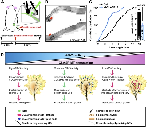
Figure 7.
CLASP1/2 are required for rapid axon regeneration in vivo. (A) Schematics of the experimental protocol for the in vivo electroporation and investigation of axon regeneration. (B) After transfection of control vector or shCLASP1 and shCLASP2 (shCLASP1/2) into the DRGs, mice were subjected to a sciatic nerve crush procedure as depicted in A. Using the whole-mount nerve segment, the lengths of all identifiable regenerating axons transfected with either control or shCLASP1/2 (encoding Venus) were measured from the crush site (marked by epineural suture, arrowheads) to the distal axon tips. Representative images of sciatic nerves (B) and quantification of axon regeneration length (five mice for each condition) (C) are shown. Bar, 1 mm. (D) A working model for GSK3–CLASP–MT triad in the regulation of axon growth. A detailed description of the model is discussed in the text.

