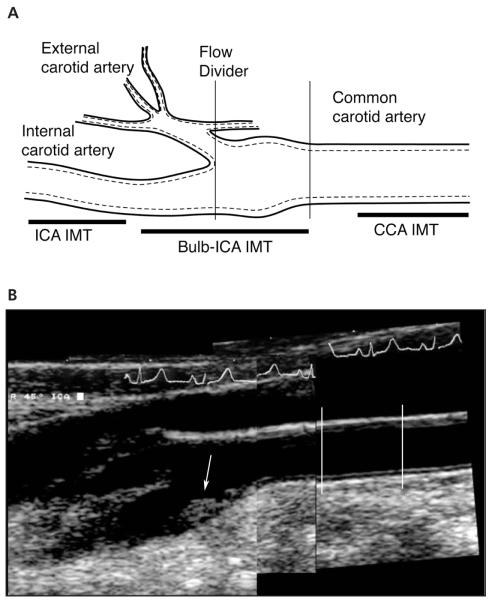
Figure 1.
A, Three levels used in the Framingham carotid IMT protocol. The CCA image is taken just before the carotid artery bulb (≈5 mm). The carotid artery bulb is measured at the level of the proximal ICA sinus, typically centered on the flow divider. The ICA measurement is made in the ICA where the walls are again parallel (ICA IMT). B, Composite carotid sonograms used as part of the Framingham IMT protocol. The CCA IMT is measured between 5 and 15 mm before the carotid bulb (lines), whereas the maximum IMT is measured at the site of the thickest wall in the carotid artery bulb or proximal ICA IMT (arrow).

