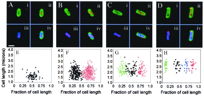
Figure 1.
Subcellular localization of RK2 in E. coli by FISH. (A–D) Strain JP704 was grown in M63/glucose at 37°C, fixed with paraformaldehyde, and hybridized with a Cy3-labeled RK2 probe. RK2 foci are red (ii, iii, and iv), the cell membranes are green because of staining with Mitotracker Green FM (i, ii, and iv), and DNA is stained with DAPI (iii and iv). Fluorescence micrographs of representative cells containing one focus (A), two foci (B), three foci (C), and four foci (D). (Bar = 1 μm.) (E–H) Subcellular distribution of RK2 in strain JP704 grown in M63/glucose at 37°C as determined by FISH. The position of each focus was measured with respect to one end of the cell. Cell length (in microns) is plotted against position of foci (given as a fraction of cell length). A total of 373 cells were measured. Graphs contain data for cells containing one (E, 18%), two (F, 60%), three (G, 14%), and four (H, 6%) foci.

