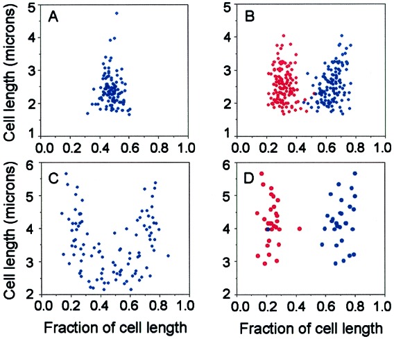
Figure 3.

Subcellular distribution of GFP-tagged pZZ6 (RK2-lacO) and pAFS52 (pUC-lacO) in E. coli. Cell length (in microns) is plotted against position of GFP foci (given as a fraction of cell length). (A and B) Strain JP872 containing RK2-lacO was grown in M63/glucose at 30°C. A total of 282 cells were measured; 39% had a single focus (A), 46% had two foci (B), and 8%, 4%, and 2% had three, four, and five foci, respectively (data not shown). (C and D) Strain JP839 containing plasmid pAFS52 was grown in LB/glucose at 23°C. A total of 124 cells were measured; 99 cells contained one focus (C) and 25 cells contained two foci (D).
