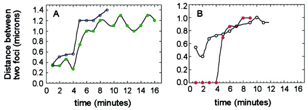
Figure 6.

Rate of separation of GFP-tagged RK2-lacO plasmids. Time-lapse microscopy of strain JP872 grown in LB/glucose at 30°C was performed as described in Materials and Methods. Images were collected 1 min apart, and the distance between foci was measured and plotted against time (min). (A) Two foci showing rapid separation over a 1-min (open blue circles) or 2-min (filled green circles) time interval. (B) Two foci showing gradual separation (open circles) and duplication of a single focus into two foci which then separate more than 0.7 μm over a 1-min time interval (filled red circles).
