Figure 1.
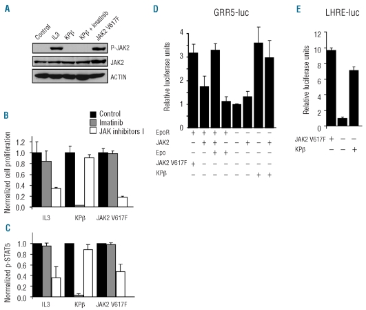
JAK2 is not required for KPβ-induced cell proliferation or STAT activation. (A) Lysates of Ba/F3-KPβ or Ba/F3-JAK2-V617F cells that were cultured in the absence of IL3 were analyzed by western blot with anti-phospho-JAK2, anti-JAK2 and anti-β-actin antibodies. As a control, Ba/F3 cells transduced with the empty vector were stimulated with IL3 or left untreated (left lane). (B) The same cells were cultured in the presence of 25 nM imatinib or 0.5 μM Jak inhibitor I for 24 h. Cell proliferation was analyzed by measuring [3H]thymidine incorporation. Untreated cells were used as a reference. Total radioactivity incorporation in the DNA of Ba/F3 cells treated with IL3, Ba/F3-KPβ and Ba/F3-JAK2-V617F was 85543 ±2026 cpm, 59098 ±1577 cpm and 44899 ±4846 cpm, respectively. (C) STAT5 phosphorylation was monitored by flow cytometry using cells treated with 0.5 μM imatinib or 2 μM JAK inhibitor I for 4 h. (D, E) JAK2-deficient γ-2A cells were co-transfected with the erythropoietin receptor (EpoR), wild-type JAK2, JAK2-V617F, KPβ or the empty vector, as indicated. Cells were co-transfected with STAT5 and the luciferase reporter pGRR5-luc (D) or pLHRE-luc (E). Cells transfected with EpoR were stimulated by erythropoietin as indicated. STAT-dependent transcriptional activities were measured and normalized using the empty vector control as the reference. One representative experiment is shown with the standard deviation calculated from triplicate measurements.

