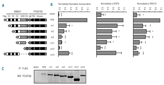
Figure 3.
KPβ coiled coils play an important role in Ba/F3 proliferation and signaling. (A) A schematic representation of KPβ and mutants is shown. Ba/F3 cells were transduced with KPβ, one of the mutants, or the empty vector as control. CC: coiled-coil domain; KOD: KANK1 oligomerization domain; Ig5: Ig-like domain 5 of PDGFRβ; TM: transmembrane domain; Kinase: split kinase domain. (B) Cells were grown for 72 h in the absence of IL3 and proliferation was measured by [3H]thymidine incorporation. Ba/F3-KPβ cells were used as a reference. All cell lines proliferated to a similar extent in the presence of IL3 (data not shown). To quantify STAT5 and ERK1/2 phosphorylation, transduced cells were washed and cultured for 4 h without IL3. Cells were permeabilized, stained with antibodies directed against phospho-STAT5 or phospho–ERK1/2 and analyzed by flow cytometry. (C) Cell lysates were immunoprecipitated overnight with 3.3 μg of FLAG antibody at 4°C to capture KPβ or mutant proteins. Antibody complexes were collected by adding protein-A/G magnetic beads for 1 h at 4°C, washed extensively and analyzed by western blot with anti-PDGFR antibodies.

